anatomical human teaching skeleton
https://mhc.andornot.com/en/permalink/artifact5074
- Dates
- 1940
- 1970
- circa 1940-1970
- Collection
- Rockwood / Kingston Psychiatric Hospital / Providence Continuing Care Centre Collection
- Category
- Anatomy
- Human Remains
- Education
- Classification
- Anatomy
- Human Remains
- Skeleton
- Bones
- Skull
- Education
- Teaching aids
- Accession Number
- 000003315
- Description
- Teaching skeleton, which consists of a human female skeleton, wired together by a central metal rod that runs down through th top of the skull, down the centre of the spinal column, and bolted in place at the bottom of the spine in the centre of the pelvis; an oval-shaped, bent wire is attached to …
5 images
- Accession Number
- 000003315
- Category
- Anatomy
- Human Remains
- Education
- Classification
- Anatomy
- Human Remains
- Skeleton
- Bones
- Skull
- Education
- Teaching aids
- MeSH Heading
- Skeleton -- models
- Anatomy -- models
- Human Remains
- Skull -- models
- Anatomy
- Skeleton
- Bones
- Skull
- Education
- Description
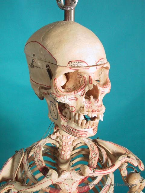
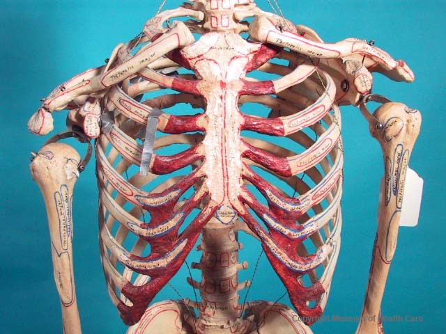
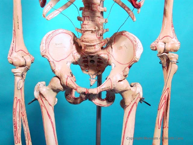
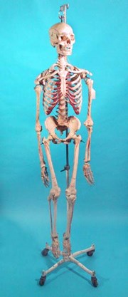
- Teaching skeleton, which consists of a human female skeleton, wired together by a central metal rod that runs down through th top of the skull, down the centre of the spinal column, and bolted in place at the bottom of the spine in the centre of the pelvis; an oval-shaped, bent wire is attached to the top of the rod to secure the skeleton to a hood; the smaller joints are joined with spring connectors; the hips are joined with bolts, wingnuts and washers; four metal wires connect the ribs; a cylindrical bar, a bolt and a square housing secure each knee joint; there is a spring on either side of the jawbone that allow it to move; on either side of the skull, there is a hook and a small bolt that secure the top of the skull, which has been cut in an oval shape, and the main part of the skull; there are several small pins set along the edge of the top piece that fit into holes along the rim of the bottom piece; the hooks can be released to allow lifting of the top of the skull to reveal the inner structure of the skull; the skeleton seems to have been covered with a varnish; the front of the rib cage has been painted a reddish brown.
- The names of the muscles and their respective insertions are written on the skeleton in black marker or paint; areas of muscles are indicated by a red outline, and their insertions in blue; all seem to be hand-painted or handwritten, including a black manufacturer's mark, which is inside a flattish diamond shape.
- Arrived hanging on irrigation stand 000.003.314
- Number Of Parts
- 1
- Provenance
- Used at the Kingston Psychiatric Hospital,
- Maker
- Denoyer-Geppert Co.
- Site Made (City)
- Chicago
- Site Made (State)
- Illinois
- Site Made (Country)
- United States of America
- Dates
- 1940
- 1970
- circa 1940-1970
- Material
- bone: yellow-white
- varnish: clear
- paint: red-brown; blue; yellow
- metal: silver
- tape: silver; cream
- Inscriptions
- "made by // Denoyer-Geppart Co. // Chicago, Illinois"
- Permanent Location
- Storage Room 0009
- 0009-2
- Temporary Location
- On exhibit “The Century: Medical Innovations of the 1800s” at Museum of Health Care, 25 June 2017.
- Dimension Notes
- Length 75.0 cm x Width 37.5 cm x Depth 23.0 cm
- Condition Remarks
- The artifact was on display in the North Gallery Room 1013 of the museum as part of the “Electricity and the Invisible Ray” from Oct. 2006 to March 2016. The skeleton was hanging on an irrigation stand 000.003.3140. There was dust and dirt all over the skeleton. There were numerous abrasions to bone surfaces caused by friction and by tangling bones hitting against each other and against the metal rod of the stand. There were several breaks and cracks on ribs, scapula and pelvis. A small piece from the metatarsal 5 was broken and loose but still held in place by supporting wire. There was a dislocation of the correct (anatomical) position of both scapulas. This was possibly caused by the breaking of the rib bones onto which the scapula was attached by means of metal connectors. The position of the scapulas shifted because there were no more securely attached to the thoracic bones. The weight of the arm also increased the dislocation of the scapula because it’s attached to the scapula and clavicle.
- There was moderate damage on the heels, tailbone and one vertebra from contact with the IV pole; the paint along the upper edges of the pelvis, the tailbone, and the breastplate had been worn; a hole had been punched in both shoulderblades and both were brittle and translucent; twelve teeth had been damaged in a previous accident, one of which was missing entirely; there were 14 cracks in the front rib cage; two of these had previously been bound with duct tape; there were nine fractures in the rear of the rib cage, five of which had been bound with duct tape and medical tape; the proper left knee had been damaged; a chunk of bone 4.0 x 3.5 cm is missing from the bottom of the femur (medial epicondyle), and a chunk 12.5 cm x 3.2 cm from the top of the tibia (medial condyle); a piece of the jawbone had been broken off, approximately 1.5 cm x 1.5 cm, from the left side where the insertion "temporalis" should appear; the skeleton was dusty; metal pieces seem to be in good condition, with no signs of significant corrosion.
- Copy Type
- Original
- Reference Types
- Book
- JPG
- Reference Comments
- "Aloe's Illustrated and Priced Catalogue of Superior Surgical Instruments, Physicians Supplies, and Hospital Furnishings," by the A.S. Aloe Co., 189–, p. 954 (imilar to item shown in Fig. 11277
- Exhibit History
- To display in Museum (North Gallery Room 1013): "Electricity and the Invisible Ray," Oct. 2006 - Feb 12, 2016.
- Agnes Etherington Art Centre - July 2002
Images