De Zeng ophthalmoscope
https://mhc.andornot.com/en/permalink/artifact10733
- Dates
- 1922
- 1950
- circa 1920-1950
- Collection
- Gillies Collection
- Category
- Diagnostic & Treatment Artifacts
- Physical Examination
- Vision
- Classification
- Ophthalmology
- Diagnosis, Eyes
- Vision
- Physical Examination
- Accession Number
- 004045009 a-f
- Description
- An ophthalmoscope in a case consisting of a black, plastic cylindrical battery handle (a) with a removable cap at the base for holding in the battery (b) and a piece at the top for a light bulb to screw in and a threaded area where the ophthalmoscope head (c) attaches; the head is flat with a circu…
3 images
- Accession Number
- 004045009 a-f
- Collection
- Gillies Collection
- Classification
- Ophthalmology
- Diagnosis, Eyes
- Vision
- Physical Examination
- MeSH Heading
- Ophthalmoscopy
- Physical Examination
- Ophthalmology
- Diagnosis, Eyes
- Vision
- MM= Ophthalmoscopy -- ophthalmoscope
- Description
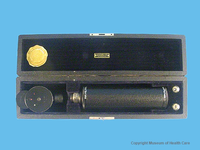
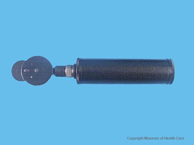
- An ophthalmoscope in a case consisting of a black, plastic cylindrical battery handle (a) with a removable cap at the base for holding in the battery (b) and a piece at the top for a light bulb to screw in and a threaded area where the ophthalmoscope head (c) attaches; the head is flat with a circular face and an adjustable dial which can be used to change the power of a lens in a hole on the face; there are two small light bulbs (d,e) and a black wooden, black velvet lined case (f).
- Number Of Parts
- 6
- Part Names
- a - handle - Size: Length 15.7 cm x Diam. 3.4 cm.
- b - cap - Size: Length 1.0 cm x Diam. 3.7 cm
- c - head - Size: Length 7.1 cm x width 4.3 cm x Depth 1.7 cm
- d - light bulb - Size: Length 2.1 cm x Diam. 1.2 cm
- e - light bulb - Size: Length 2.1 cm x Diam. 1.2 cm
- f - case - Size: Length 27.8 cm x Width 6.8 cm x Depth 5.5 cm
- Provenance
- This artifact was used by the donor's uncle Dr. Walter Barnhart during the 1920s to the 1940s.
- Maker
- De Zeng
- Site Made (City)
- St. Louis; Camden
- Site Made (State)
- Missouri; New Jersey
- Site Made (Country)
- United States of America
- Dates
- 1922
- 1950
- circa 1920-1950
- Date Remarks
- marking on the head says it was patented in 1922
- Material
- plastic: black
- wood: black
- fabric: black
- glass: clear
- metal: yellow
- Inscriptions
- "DeZeng // Simplex // No. 110" engraved on the face front; "Made in // U.S.A. // PAT. // 12.21.15 // 7.18.22" engraved on the face back; "DeZeng // OPTOMETERS // PHOROMETERS // RETINOSCOPES // OPHTHALMOSCOPES // PHARYNGOSCOPES // CONTROLERS/ PERIMETERS // OTOSCOPES // ETC. // CAMDEN, NEW JERSEY, U.S.A" on a gold metal label inside the case; "A. S. ALOE -CO // ST. LOUIS, MO." on a small metal plate in the case
- Permanent Location
- Storage Room 0010
- 0010-E4-5
- Temporary Location
- Device only: On exhibit: "Ophthalmology: The First 50 Years," at Hotel Dieu Hospital, Kingston, L-2005.8.
- Condition Remarks
- There are a few chips missing from the case; the battery is very corroded; #2: the lid of the case is very loose.
- Reference Types
- Person
- Reference Comments
- Richard Gillies
- Research Facts
- ophthalmoscope [of-thal´mo-skop] an instrument for examining the interior of the eye. Direct ophthalmoscope one that produces an upright, or unreversed, image of approximately 15 times magnification. The direct ophthalmoscope is used to inspect the fundus of the eye, which is the back portion of the interior eyeball. Examination is best carried out in a darkened room. The examiner looks for changes in the color or pigment of the fundus, changes in the caliber and shape of retinal blood vessels, and any abnormalities in the macula lutea, the portion of the retina that receives and analyzes light only from the very center of the visual field. Macular degeneration and opacities of the lens can be seen through direct ophthalmoscopy.
- The donor graduated from medical school in 1955.
Images