anatomical female model
https://mhc.andornot.com/en/permalink/artifact5284
- Dates
- 1943
- circa 1943
- Collection
- Rockwood / Kingston Psychiatric Hospital / Providence Continuing Care Centre Collection
- Category
- Anatomy
- Education
- Physiology
- Classification
- Anatomy
- Education
- Physiology
- Accession Number
- 000003347 a-l
- Description
- Female teaching model, which consists of a painted plaster figure mounted on a wooden base with multiple removable parts, detailing all of the body's major anatomical parts; the main teaching model (a) consists of a plaster female form from mid-thigh upwards, minus the arms past the shoulders; the …
5 images
- Accession Number
- 000003347 a-l
- Category
- Anatomy
- Education
- Physiology
- Classification
- Anatomy
- Education
- Physiology
- MeSH Heading
- Anatomy -- models
- Anatomy
- Education
- Physiology
- Description
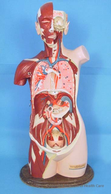
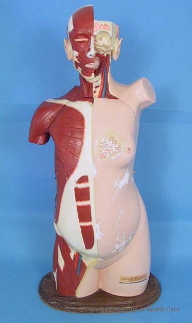
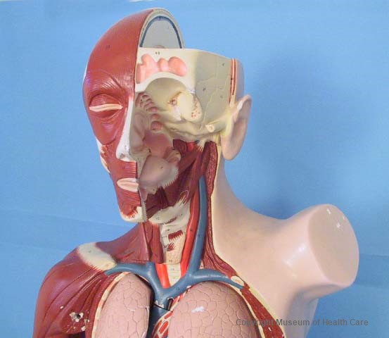
- Female teaching model, which consists of a painted plaster figure mounted on a wooden base with multiple removable parts, detailing all of the body's major anatomical parts; the main teaching model (a) consists of a plaster female form from mid-thigh upwards, minus the arms past the shoulders; the plaster model is affixed by the base of the thighs to an oval, bevelled-edged wooden base, possibly lacquered walnut; a partially removed and illegible paper label is affixed to the top surface of the base at the back; the thigh, pubic area, side, shoulder, buttocks, ear, and ¼ of the back of the head; on the proper left side of the model is shown the outer, pink, skin covering; a rectangular label is affixed to the front of the model on the thigh on the proper left side; the front of the model is cut away to reveal the body cavity, the reproductive organs, major veins and arteries, the digestive organs, the lungs, a partial view of the ribs, the pharynx, muscle tissue, a partial view of the mouth and teeth, the skull, ligaments, and vertebrae; there are two indentations on the inner bottom of the cavity to accomodate the bottom tip of parts (k) and (l), the uterus and fallopian tubes and the uterus cover; (k) consists of a triangular piece of painted plaster, its bottom rounded tip formed to resemble one-half of the vagina; a curving, rounded tube extends from each of its top two corners, to form the fallopian tubes, and at the end of the proper left tube, an ovary; the end of the proper right tube is damaged and an inner metal wire, curved at its end, is exposed; the outer surface of (k) is painted cream, its outer tips and corners are pink; its inner surface has an outer triangle of dark red with alternating blue and red dots; an orange-pink triangle runs along the inside of the dark red triangle; it also contains alternating blue and red dots; the inner centre of (k) is light pink with two metal pegs extending from its centre; these fit into two holes in the centre of (l), the uterus front; (l) is triangular, with rounded corners, its bottom tip forming one-half of the vagina; its outer surface is cream and its inner surface matches the inner surface of (k); the inner pink part of (k) and (l) represents the uterine body cavity, the orange-pink triangle possibly the endometrium, and the dark red triangle the myometrium; (j) attaches to (a) by a metal rod extending from the inner surface of (j) that fits into a hole in the centre of the body cavity below a cut-away of the small intestine; (j) depicts the large and small intestines, with the major veins and arteries supplying blood to the area on the back of the part; the appendix is represented by a black rubber peg extending from the back, lower proper left corner; at the bottom proper right-hand corner of the front of the intestines, the caecum is depicted as a rounded endpiece on a hinge; this can be lifted to reveal the ileocaecal valve and a pink, ridged surface; (i) one kidney attaches by a metal peg extending from its back side into a hole in the body cavity to the proper left of the intestines; its front surface is dark brown with cream-coloured detail along its concave edge; its back surface shows blue and red capillaries, the medulla, cortex, and calyx; to the proper left of the removable kidney, and above the intestines, (h) the stomach attaches to the body cavity by a metal peg extending from the s tomach's top proper right rear corner, inserted into a small hole in the centre of the cavity above and to the proper right of the hole for the intestines; the stomach (h) is largely cream-coloured, with red lines detailing its blood supply on its front, and a brown circular area in each top corner illustratin the gastroesophageal opening and possibly the duodenal bulb; above the intestines, (j) the liver attaches to the cavity by a metal peg that extends from its rear top surface, and fits into a hole above the depiction of what appears to be the gastroesophageal opening in the cavity; the liver is a deep red colour on its front surface; the model shows the inferior vena cava running from the top of the liver to its rear surface; the rear surface also shows the gall bladder, in dark green with the common hepatic duct, the hepatic portal vein in purple, and the hepatic artery in bright red; the round ligament is also illustrated as a cream-coloured line; above the liver, in the body cavity, (e) and (f), the front and back of the heart, attach by a bent metal peg that extends from the top of the back of the heart and is inserted into a hole in the body cavity between the cut-aways of the carotid artery and the superior vena cava; the back of the heart (f) shows the outer surface of the left ventricle and right ventricle, the coronary sinus, posterior interventricular branch of the right coronary artery, the middle coronary vein, the posterior artery and vein of the left ventricle, the posterior interventricular suculus, the inferior vena cava, the right and left pulmonary veins, and the left and right pulmonary arteries; the inner surface of the back of the heart illustrates the right atrium, the aorta and the aortic semilunar valve, and the left ventricle; the right and left AV valves and the chordae tendineae are depicted by two loops of cream-coloured fabric, smooth along their top edges and with triangular points running along their bottom edges; cream-painted wood fibres extend from these fabric triangles and attach to two brown ridges on each side, representing papillary muscles; this half also has a ridge running down its centre representing the interventricular septum, into which a hole has been drilled approximately halfway down its length; (e) the front of the heart has a metal peg extending from its interventricular septum and this fits into the hole in (f); the front of the heart illustrates the pulmonary trunk, the aortic semilunar valve, the right atrium, right ventricle, and left ventricle in its inner surface; the outer surface of the front of the heart shows the left and right coronary arteries and veins, the auricle of the left atrium, the ligamentum arteriosum, and the auricle of the right atrium; to the proper left and right of the heart, (c) and (d) the proper left and right lungs attach into two holes in the body cavity by way of a metal peg that extends from the flat side of both lungs; both lungs are four-sided with three long sides, one flat, one convex, and one concave; the shorter, bottom sides are cut at a curving approximately 20° angle; the convex and concave sides of the lungs are pink, with grooved patches covering the surface to represent the outer surface of the lung; both bottom sides are triangular, with the long edge detailed in dark red and the remainder a cream colour; the flat side of the proper left lung is pink with red and blue dots with dark red centres and cream dots with pink centres; the flat side of (d), the proper right lung, has cream-coloured, branchlike details extending from the concave edge as well as five sets of blue and red dots with dark red centres; the bottom tip of (d) is cut on a straight angle; the remainder of (a), the main teaching dummy, consists of a neck cutaway in the front to reveal musculature, the trachea, major arteries and veins; the face is cut away on one side to reveal the structure of the mouth, sinus cavities, eye socket and skull; the top of this proper left side is cut away to show strata of the skull; the proper right side of the face and skull detail musculature in dark red and bone in cream; both sides of the head have extending, full ears; the back of the head from the centre to proper right, and extending to the lower back, is cut away; vertebrae, musculature, nerves, the back of the proper right lung are detailed; a section of the lumbar vertebrae is missing, with two triangular pieces of plastic set into the top and bottom of the hole in the spinal column; on either side of the model, approximately at the halfway point near the front, there is a small hole drilled in the plaster; the hole on the proper left side is empty, while the one on the proper right has a metal hook latch attached with a screw; another hole is drilled into the plaster in each of the model's armpits; these holes accomodate two metal pegs extending from each side of the top end of (b), the body cavity front; at the approximate midpoint of the cover, on both outer surface edges, there is a metal loop set into the plaster; the proper right loop accomodates the hook latch on the proper right side of the model; the body cavity front is oval with a convex outer surface and a concave inner surface; its inner surface illustrates the position of the ribs, sternum, muscles between the ribs (intercostals), arteries and veins, abdominal muscles, and umbilical ligaments; the outer edge of the cover shows layers of dermis, muscle and bone; the front outer surface of the cover is half cut away, so that the proper left side illustrates the outer skin surface, and a cut away breast revealing the alveoli; the proper right side illustrates the positions of the rectus abdominus, pectoralis major, serratus anterior, and the external abdominal oblique, all in dark red with a ridged texture.
- Number Of Parts
- 12
- Part Names
- a - teaching model - Size: Length 94.0 cm x Width 39.0 cm x Depth 22.5 cm
- b - body cavity, front - Size: Length 51.5 cm x Width 32.7 cm x Depth 10.5 cm
- c - proper left lung - Size: Length 19.3 cm x Width 10.2 cm x Depth 5.6 cm
- d - proper right lung - Size: Length 20.0 cm x Width 13.1 cm x Depth 5.5 cm
- e - heart, front - Size: Length 12.5 cm x Width 10.5 cm x Depth 4.7 cm
- f - heart, back - Size: Length 15.0 cm x Width 9.0 cm x Depth 7.0 cm
- g - liver - Size: Length 21.5 cm x Width 13.5 cm x Depth 11.0 cm
- h - stomach - Size: Length 15.0 cm x Width 11.0 cm x Depth 9.0 cm
- i - kidney - Size: Length 8.0 cm x Width 4.4 cm x Depth 3.3 cm
- j - intestines - Size: Length 23.0 cm x Width 22.0 cm x Depth 14.5 cm
- k - uterus and fallopian tubes - Size: Length 11.0 cm x Width 10.5 cm x Depth 6.0 cm
- l - uterus, front - Size: Length 8.5 cm x Width 5.2 cm x Depth 1.5 cm
- Provenance
- Used at the Kingston Psychiatric Hospital
- Maker
- Denoyer-Geppert Company
- Site Made (City)
- Chicago
- Site Made (State)
- Illinois
- Site Made (Country)
- United States of America
- Dates
- 1943
- circa 1943
- Material
- metal: brown-yellow
- wood: brown
- plaster: white
- paint: pink; white; red; blue; dark red; turquoise; dark green; brown; purple; yellow
- paper: yellow; blue; white; black
- rubber: black
- fabric: cream
- Inscriptions
- (a) Imprinted into the plaster surface of the back of the model's proper left thigh: "Made in U.S.A. // Denoyer-Geppert Co. // Chicago Ill. 1943"; below and to the proper right of this is, "Br26 '71" written in pencil; the rectangular label on the front of the proper left thigh reads, "PRODUCED BY // DENOYER-GEPPERT COMPANY // CHICAGO, ILLINOIS // MADE IN U.S.A."; on the model and all its corresponding pieces, there are numbers handwritten in black marker referring to specific anatomical parts; on the front and back surfaces of (a), the numbers extend from the base to the top of the head (see accession sheet for numbers); on the top surface where the skull is cut away the numbers extend from top to bottom.
- Permanent Location
- Storage Room 0010
- 0010-F5-5
- Condition Remarks
- The outer parts were dusty; the wooden base of (a) was in good condition, with minor varnish wear; the proper left side of (a), which was mostly smooth and painted with a light flesh tone, was in poor condition; 90% of this side showed cracks in the top painted layer; this was possibly the result of the paint being unable to bond to the smooth plastic surface; the paint on this side was flaking off in patches; major damage included a patch revealing the inner plaster surface at the back of the model near the base, 2.1 cm x 1.3 cm; a pronounced web of hairline cracks in the paint had developed underneath the manufacturer's information; another flaked patch appears near the base on the back side near the proper left side, 4.0 cm x 2.3 cm; there were 13 smaller patches on the lower portion of the model; a chip of paint had come away from the area surrounding the hole at the midpoint of the proper left side that partially secured (b); the plaster in this area showed signs of moderate use; underneath the shoulder on this side a chip of paint 2.0 cm x 1.5 cm had come away from the plaster; there were approximately 12 smaller chips in the paint in this area; at the top of the shoulder, there were three chips in the paint, the largest 0.5 cm x 0.8 cm; next to these was a hole in the plaster, 0.6 cm x 0.4 cm; there was a nick in the ear of this proper left side, 0.5 cm x 1.0 cm that appeared to be as a result of use and handling; there was a minor chip in the plaster on the top proper left side of the skull, near the top rim of the cut-away portion; the flesh-toned areas all show evidence of handling in the form of a grey film of fingerprints; at the front of the model in the bottom proper left corner there were eight minor patches of revealed plaster; there was a major patch at the bottom centre of the model on the rim of the cut-away front piece, 2.5 cm x 0.5 cm; there were approximately five minor patches of exposed plaster in the lower proper right corner of the model; on the surface of the bottom centre of the cavity there were three minor patches of exposed plaster, the largest of which was 0.5 cm x 0.7 cm; on the lower bend of the large intestine, there was a patch of exposed plaster 2.0 cm x 0.9 cm; the paint surrounding each of the seven organ attachment holes was worn, as were the two holes used to attach the body cavity front (b); the paint was significantly worn from the ends of the carotid artery that comes in contact with the proper right side of the heart; there were several worn patches on the carotic artery and superior vena cava that run across the top of the body cavity from shoulder to shoulder; there were six major patches, and from proper right to left they were 0.5 cm x 0.3 cm, 1.0 cm x 0.4 cm, 0.6 cm x 0.3 cm, 1.4 cm x 0.5 cm, 1.2 cm x 0.4 cm, and 0.5 cm x 0.4 cm; there was a patch of black discolouration on the chin and nose, 5.0 cm x 1.0 cm and 1.7 cm x 1.0 cm; there was a small chip on the lips, as well as the tip of the nose, and the corner of the proper left eye socket; in the proper left eye socket there was a hole that appeared to once accomodate another part; there was adhesive residue surrounding it from previous repairs; and a worn patch 1.5 cm x 1.0 cm; the ridges of the vertebrae on the back side were dirty; there was a significant chip 1.5 cm x 0.5 cm on the proper left edge of the cut-away vertebrae section; there was a chip 0.7 cm x 0.3 cm in the 10th vertebra from the top on its proper left side, and another chip in the lowest vertebra 1.0 cm x 0.4 cm; on the back side of the head, on the proper right side depicting the skull, there was a shallow gouge in the paint and plaster 1.5 cm x 3.0 cm; (b) the body cavity front was in poor condition; there were two hairline cracks in the plaster running from its top end to its approximate midpoint; the area surrounding the proper right metal connecting loop was cracked, and there were plaster chunks missing; the metal wire originally encased in the plaster that emerges to form the loop was visiboe; a fibrous material was also revealed due to the damage; this was probably the result of strain caused by the hook on the opposite side of (a) being missing, so the proper left metal ring bears the majority of the cover's weight; there were additional cracks in the lower portion of the cover radiating from its bottom edge (approximately 8 in number); along the edges of the cover there were chips and flakes in the paint and plaster; below the proper right hook there were approximately 19 chips in the plaster, the largest of which was 4.0 cm x 0.4 cm; along the proper left edge there were approximately 15 chips, the largest of which was 3.5 cm x 0.8 cm; on the inner surface of the cover there was a chip in the plaster 3.2 cm x 2.5 cm; both attachment pegs were slightly bent; on the proper left side of the front surface, where the skin is depicted, approximately 75% of the paint shows signs of flaking and deterioration; there is a major patch of flaking in the bottom proper left corner, where the plaster is revealed, approximately 12.5 cm x 16.0 cm; there is a gouge in the plaster below the proper left breast, approximately 12.0 cm x 6.0 cm; there were two smaller gouges in the plaster on the bottom edge of the cut-away portion of the proper left breast, one 1.0 cm x 0.6 cm and the other 1.1 cm x 0.4 cm; along the centre ridge of the cover's top surface there were two gouges in the plaster, the lower 5.8 cm x 2.9 cm and the upper 4.2 cm x 1.2 cm; there were hairline cracks similar to those on the bottom surface running throughout the plaster; on the proper right side of the cover at the midpoint, there were two brown marks, the lower 0.5 cm x 0.7 cm and the upper 0.2 cm x 0.5 cm; at the midpoint of the bottom edge, there was another gouge in the plaster 1.7 cm x 2.4 cm; brown fibres are visible in the plaster in this gouge; the proper left lung (c) was in good condition; there was a chip in the paint 1.2 cm x 0.6 cm on its outer surface in the bottom proper left corner; as well as 12 minor chips in the top proper right corner; there were two minor chips in the top proper right corner of the long concave surface, and one significant chip in the paint and plaster 1.4 cm x 0.5 cm in the lower proper left corner; the metal peg showed no signs of corrosion; there were three minor chips in the paint in the proper right corner of the top short side, and one minor chip in the proper right corner; the proper right lung (d) was in fair condition, with significant damage in the proper right corner of the top short concave end, where there was a series of chips in the plaster in an area 3.2 cm x 2.5 cm; and below this along the edge of the longer concave side, where there was a hole in the object surrounded by chips in the plaster 3.7 cm x 1.0 cm; there were approximately 16 minor chips in the paint on the large convex surface; the metal peg showed no signs of corrosion; the front of the heart (e) was in fair condition; its inner surface had approximately 9 minor chips in the paint, and the outer surface had a hole in the plaster 0.7 cm in diameter in the top proper right corner; there were also several hairline cracks in this surface, suggesting possible structural damage; beneath the hole there was a chip in the paint 1.0 cm x 0.4 cm; in the lower proper left corner of the outer surface there were five chips in the paint: 0.8 x 0.2 cm, 0.4 cm x 0.3 cm, 0.6 cm x 0.2 cm, 0.3 cm x 0.6 cm, and 0.2 cm x 0.1 cm; the metal peg showed no sign of corrosion; the back of the heart (f) was in good condition; there was some minor chipping of the paint along the edges of the ends of the arteries and veins, and approximately 17 minor chips in the paint on its front surface; there were two blue smudges on its pink surface, likely fingerprints; there were no signs of corrosion on the metal peg; on the back surface there were approximately eight minor chips in the paint; there were pencil marks on the inner surfaces of the left and right atrium; the liver (g) was in poor condition; there were cracks in approximately 10% of the surface; there were chunks missing from the plaster on the large convex surface: 7.5 cm x 3.2 cm, 3.3 cm x 3.4 cm, 1.5 cm x 1.4 cm; there were chunks missing from the shorter, slightly concave, end: 1.4 cm x 0.6 cm, 3.8 cm x 1.5 cm, 0.6 cm x 1.4 cm; there were chips in the plaster along 90% of the rim of the large concave side; the metal peg showed no signs of corrosion; (h) the stomach was in good condition; its surface was slightly dirty, with faint smudges that appeared to be fingerprints; the metal peg showed no signs of corrosion; there was a chip in the plaster and paint 1.5 cm x 0.6 cm in the corner without the metal peg; there were no other cracks or chips; (i) the kidney was in good condition; its outer surface had one chip in the paint 0.4 cm x 0.2 cm in the top half, and one chip in the paint and plaster in the lower proper left corner 0.7 cm x 0.8 cm; there were approximately 10 minor chips along the kidneys' edge, and one significant chip in the plaster in the inner surface in the top proper left corner 0.6 cm x 0.4 cm; (j) the intestines were in good condition; the cream-coloured surfaces showed evidence of fingerprints; the lower end of the large intestines had chips in the paint on the inner surface; the top one was 0.4 cm x 0.5 cm, below this was a chip 3.4 cm x 0.4 cm, below this was 0.3 cm x 0.3 cm, and the lowest chip in the plaster was 0.6 cm x 0.5 cm; there were several smaller chips along the edge of the end of the lower intestines; there were two chips in the paint on the front surface of the intestines, one in the top proper left corner, 0.6 cm x 0.8 cm, and the other 0.7 cm x 0.4 cm, along the edge of the hinged part representing the caecum; there were no other cracks or chips, and the metal peg showed no signs of corrosion; (k) the uterus back with the fallopian tubes was in fair condition; the proper left fallopian tube had broken and the inner wire was exposed; there was a yellowing adhesive on this surface, suggesting past repairs; the inner surface of the uterus had a chip in the paint and plaster 1.6 cm x 0.4 cm in the proper left corner, and another minor chip in the paint surrounding the proper left peg; the proper right fallopian tube had structural cracks bisecting its base at the proper right corner of the uterus; there was a blue pen mark in the centre of the inner surface of the uterus; on the outer surface of (k), there was a chunk of plaster missing from the base of each fallopian tube; the one on the proper right was 1.7 cm x 1.4 cm, and on the proper left, 1.1 cm x 1.5 cm; in the proper right corner the end of one of the inner supporting wires had forced its way through the plaster; in the lower half of the uterus a faint crack ran across its length; the bottom of the vagina had a faint, dark discolouration, from handling or repeated contact with (b); (l) the front of the uterus was in poor condition; 75% of its edges were chipped and cracked; two large chunks of plaster were missing from the middle of its inner surface: 1.5 cm x 1.1 cm on the proper left side and 0.8 cm x 0.7 cm on the proper right side; on the inner surface there was a crack running from the midpoint of the proper left side to the top proper left corner; the front surface was slightly dirty; there were three significant cracks bisecting its width and one crack running from its top end to its midpoint.
- Copy Type
- Original
- Reference Types
- Person
- Photos
- Reference Comments
- Dr. James Low
- Research Facts
- Probably used for education of nurses, in oral and written examinations and demonstrations.
- Exhibit History
- May 2006: "Medical Art Through the Ages: The History of Anatomical Teaching Models."
Images