Varicose ulcers [anatomical model]
https://mhc.andornot.com/en/permalink/artifact8406
- Dates
- 1934
- circa 1934
- Collection
- University Health Network - Academy of Medicine Collection
- Category
- Anatomy
- Art
- Moulage
- Classification
- Anatomy -- models
- Anatomy
- Art
- Moulage
- Accession Number
- 1985.8.2
- Description
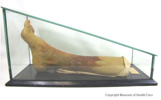
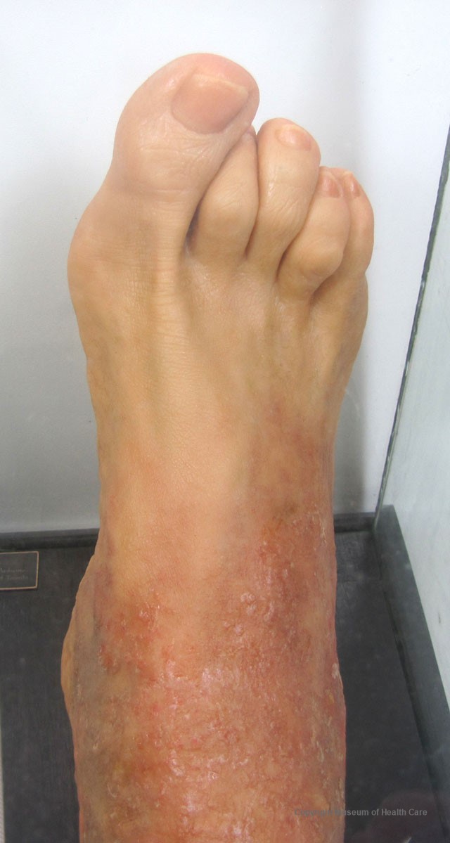
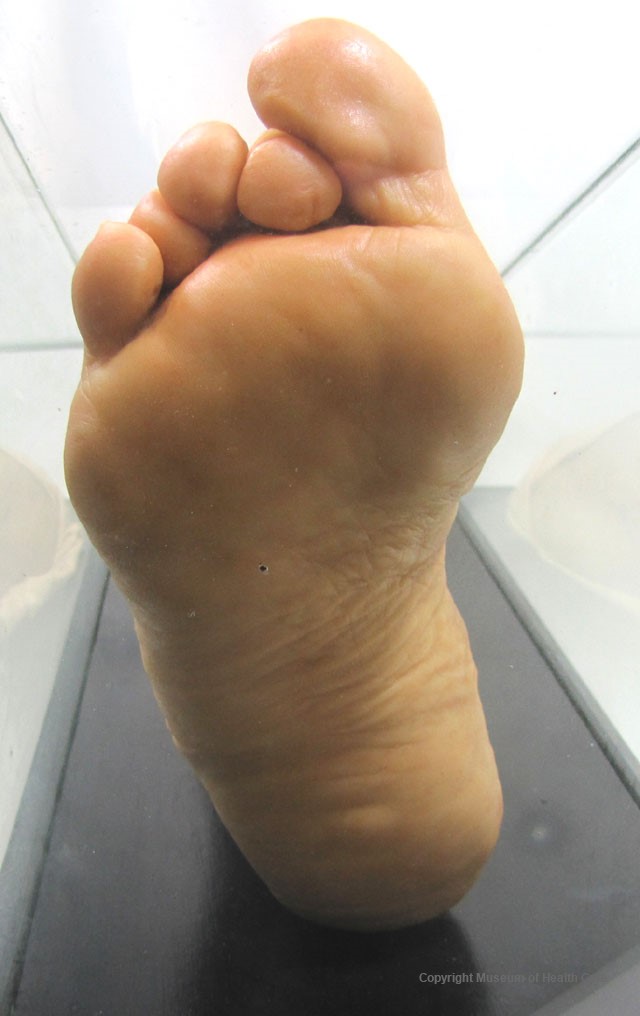
- A wax model of the right leg below the knee showing varicose ulcers; in a glass case mounted on a wooden base; signed by maker; has a label identifying the model; very heavy.
5 images
- Accession Number
- 1985.8.2
- Classification
- Anatomy -- models
- Anatomy
- Art
- Moulage
- Description
- A wax model of the right leg below the knee showing varicose ulcers; in a glass case mounted on a wooden base; signed by maker; has a label identifying the model; very heavy.
- Number Of Parts
- 1
- Provenance
- Acquired from the Academy of Medicine; source: Professor N. Joy, Art as Applied to Medicine, University of Toronto.
- Maker
- Maria T. Wishart and D. I. Foster
- Site Made (City)
- Toronto
- Site Made (State)
- Ontario
- Site Made (Country)
- Canada
- Dates
- 1934
- circa 1934
- Date Remarks
- Printed on label
- Material
- wax: flesh; red
- glass: clear
- wood: black
- fabric: white
- paper: white
- Inscriptions
- On label: "Mr. S. S. for Dr. W. E. Gallie, Dept. of Surgery"; signed "Wishart and Foster '34"
- Permanent Location
- Storage Room 0010
- 0010-F8-1
- Length
- 57.5 cm
- Width
- 29.0 cm
- Depth
- 23.0 cm (case)
- Unit Of Measure
- centimeters
- Condition Remarks
- June 2023 - overall excellent condition; some surface dirt and marks on glass case; flaking of black paint around edges of wooden base. Handling note: Heavy, handle with care.
- Copy Type
- Original
- Reference Types
- Other
- Reference Comments
- See also 1985.8.1 and 1982.18
- Research Facts
- Venous ulcer is defined by the American Venous Forum as "a full-thickness defect of skin, most frequently in the ankle region, that fails to heal spontaneously and is sustained by chronic venous disease, based on venous duplex ultrasound testing." Venous ulcers are wounds that are thought to occur due to improper functioning of venous valves, usually of the legs (hence leg ulcers). They are an important cause of chronic wounds, affecting 1% of the population. Venous ulcers develop mostly along the medial distal leg, and can be painful with negative effects on quality of life.
- Exercise, together with compression stockings, increases healing. The NICE guideline recommends that everyone with a venous leg ulcer, even if healed, should be referred to a vascular specialist for venous duplex ultrasound and assessment for endovenous surgery.
- M. Wishart was trained under Max Broedell at Johns Hopkins; the model was made for the Art Service, Faculty of Medicine, University of Toronto.
- Exhibit History
- May 2006: "Medical Art Through the Ages: The History of Anatomical Teaching Models."
- On exhibit: "Medical Art throughout the Ages: Moulages," Kingston Museum of Health Care, Sept. 2007
Images